39 eye diagram not labeled
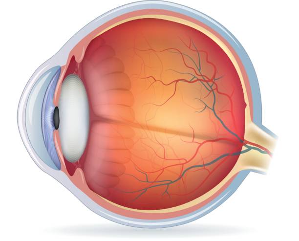
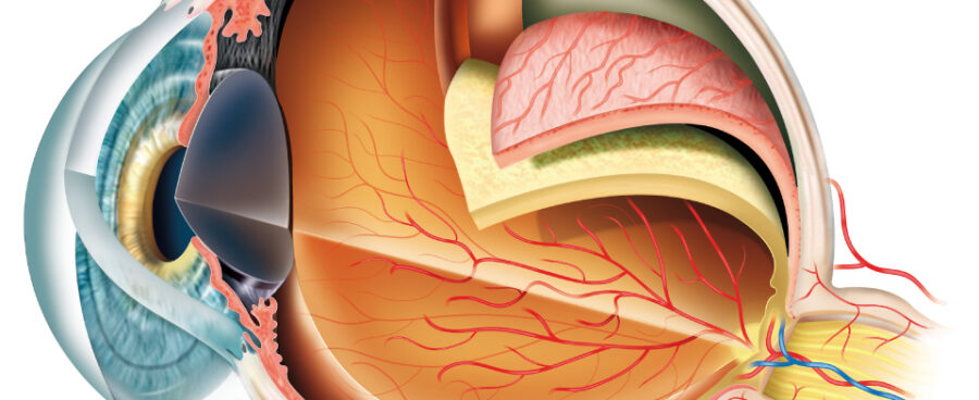
Eye Diagram Basics: Reading, Analyzing and Applying Dec 16, 2011 · Eye diagrams provide instant visual data that engineers can use to check the signal integrity of a design and uncover problems early in the design process. Used in conjunction with other measurements such as bit-error rate, an eye diagram can help a designer predict performance and identify possible sources of problems. Related articles: What Does the Eye Look Like? - Diagram of the Eye | Harvard Eye Associates Vitreous Gel: A thick, transparent liquid that fills the center of the eye. It is mostly water and gives the eye its form and shape. Our eyes are vital for seeing the world around us. Keep them healthy by maintaining regular vision exams. Contact Harvard Eye Associates at 949-951-2020 or harvardeye.com to schedule an appointment today.
16167 results for eye diagram in all - Adobe Stock Search from thousands of royalty-free Eye Diagram stock images and video for ... Eye anatomy with labeled structure scheme for human optic outline diagram.
Eye diagram not labeled
Eye Anatomy: The 9 Main Parts of the Eye | Specialty Eye Institute Optic Nerve. The second cranial nerve. The next component of the eye to be familiar with when you are analyzing eye anatomy is the optic nerve. The largest sensory nerve of the eye; carries impulses for sight from the retina to the brain. Composed of retinal nerve fibers that exit the eyeball through the optic disc, traverse the orbit, and pass ... Human Eye Diagram, How The Eye Work -15 Amazing Facts of Eye The tear gland located above each eyeball and inside your upper eyelid. This gland is responsible for making a fluid that is mostly salt and water (tears) to keep the surface of your eyeball clean and moist. It also protects your eye from damage. Also Read: Brain Facts | Blood Fact s | Respiratory System | Digestive System The Eye - Science Quiz - Seterra - GeoGuessr The anatomy of the eye is fascinating, and this quiz game will help you ... Light enters our eyes through the pupil, then passes through a lens and the ...
Eye diagram not labeled. Human eye | Definition, Anatomy, Diagram, Function, & Facts Apr 19, 2023 ... human eye, in humans, specialized sense organ capable of ... Opening of the eye is not just the result of passive relaxation of the ... Parts of the Eye | National Eye Institute Check out this fact sheet to see a labeled diagram of the eye and learn about the different parts of the eye. Topics: Glaucoma. Material Type: Handouts. Download. English: Parts of the Eye (PDF 603.5 KB) Spanish: Las partes del ojo (PDF 897.7 KB) National Eye Institute. Facebook; Twitter; LinkedIn; Cow's Eye Dissection | Exploratorium Step 2: Muscles move the eye Without moving your head, look up. Look down. Look all around. Six muscles attached to your eyeball move your eye so you can look in different directions. Cows have only four muscles that control their eyes. They can look up, down, left, and right, but they can't roll their eyes like you can. Eye anatomy: Muscles, arteries, nerves and lacrimal gland ... Apr 12, 2023 · Orbit definition. Bony cavity within the skull that houses the eye and its associated structures (muscles of the eye, eyelid, periorbital fat, lacrimal apparatus) Bones of the orbit. Maxilla, zygomatic bone, frontal bone, ethmoid bone, lacrimal bone, sphenoid bone and palatine bone. Structure of the eye. Cornea, anterior chamber, lens, vitreous ...
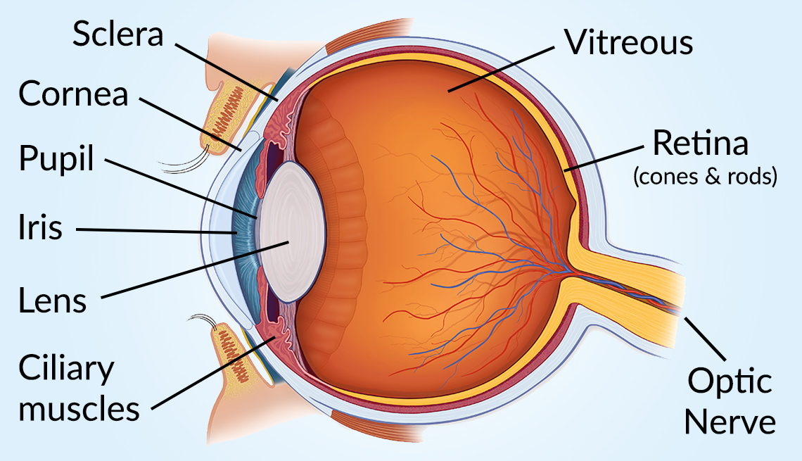
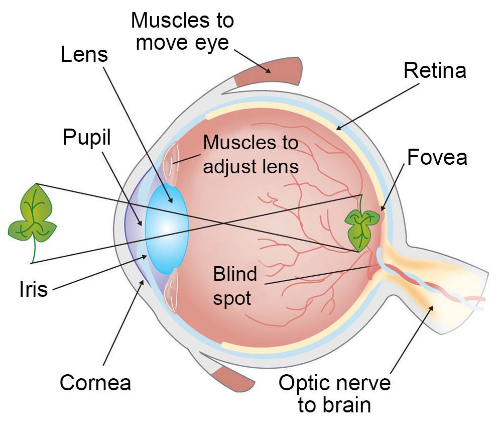
PDF Parts of the Eye - National Eye Institute Iris: The iris is the colored part of the eye that regulates the amount of light entering the eye. Lens: The lens is a clear part of the eye behind the iris that helps to focus light, or an image, on the retina. Macula: The macula is the small, sensitive area of the retina that gives central vision. It is located in the center of the retina. Lacrimal Gland - American Academy of Ophthalmology Glándula Lagrimal. Mar. 28, 2016. Located above the eye, this structure produces tears. Tear Drainage System. Read an overview of general eye anatomy to learn how the parts of the eye work together. International Education Award. Commitment to Advocacy Award. Module 1: Labeled Diagram of the Eye - Pinterest Labeled Diagram of the Eye Diagram Of The Eye, Eye Anatomy, Visual System,. More like this ... Simple eye diagrams | Easy eye diagram | Labeled eye diagram ... Structure and Function of the Eyes - Eye Disorders - MSD Manuals Rods are more numerous than cones and much more sensitive to light, but they do not register color or contribute to detailed central vision as the cones do.
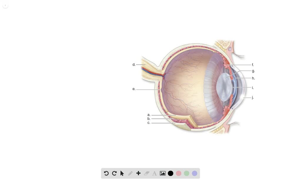
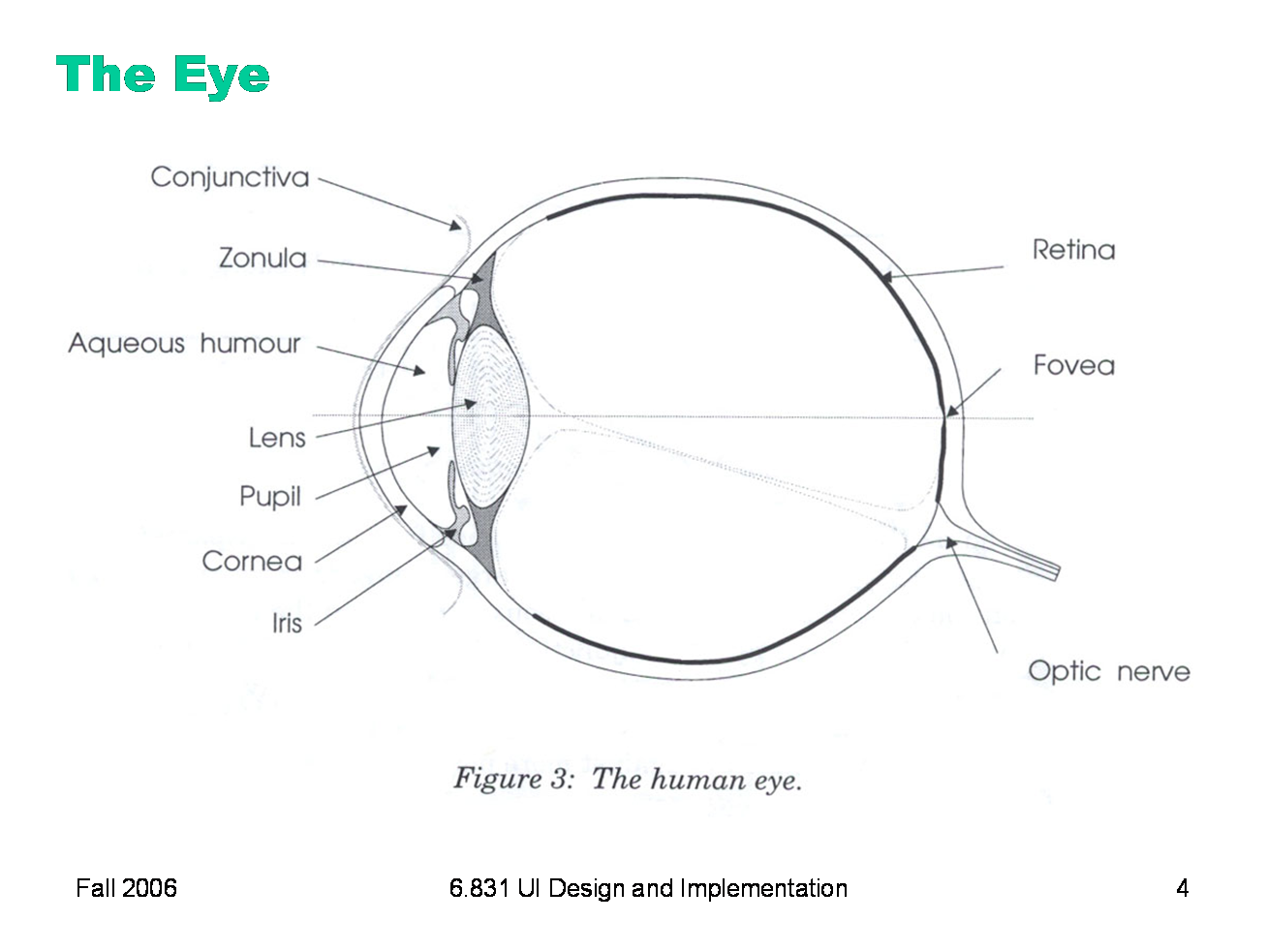
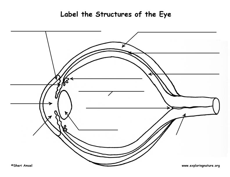
Labelling the eye — Science Learning Hub Drag and drop the text labels onto the boxes next to the eye diagram If you want to redo an answer, click on the box and the answer will go back to the top so you can move it to another box. If you want to check your answers, use the 'Reset incorrect' button. This will reset incorrect answers only. The structure of the eye (video) | Khan Academy In this video, we're going to talk about the structure of the eye. And we're going to do that by drawing a cross-sectional diagram of the eyeball. The first thing we're going to draw is the white part of the eye, which is known as the sclera. So I'm just drawing that in. And I'm going to label is sclera. Anatomy of the Eye | Johns Hopkins Medicine Ciliary body. The part of the eye that produces aqueous humor. Cornea. The clear, dome-shaped surface that covers the front of the eye. Iris. The colored part of the eye. The iris is partly responsible for regulating the amount of light permitted to enter the eye. Lens (also called crystalline lens). Anatomy of the eye: Quizzes and diagrams | Kenhub Sep 14, 2022 · Unlabeled diagram of the eye Click below to download our free unlabeled diagram of the eye. See how many of the blanks your memory allows you to fill in, then check your answers against the labeled diagram. Download PDF Worksheet (blank) Download PDF Worksheet (labeled) How did you do?
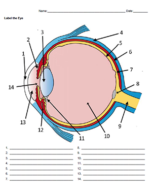
Label the Eye
Diagram of the Eye - Lions Eye Institute To understand the eye and its functions, it's important to understand how the eye works, see below diagrams for both the external eye and the internal eye.

Module 1: Labeled Diagram of the Eye | Diagram of the eye ...
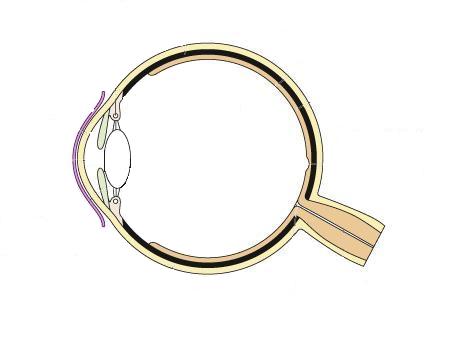
Label the Eye - The Biology Corner The image was modified from an eye diagram at Wikimedia Commons. I added the numbers and additional errors to identify structures that weren't on the original diagram. There are a few terms that can be vague, for example, the aqueous humor could also be labeled as the aqueous chamber. Zonule of Zinn can also be called suspensory ligaments.

Choroid - Wikipedia
The Eyes (Human Anatomy): Diagram, Optic Nerve, Iris ... - WebMD The weaker eye, which may or may not wander, is called the "lazy eye." Astigmatism: A problem with the curve of your cornea. If you have it, your eye can’t focus light onto the retina the...

Q10 Draw a labeled sketch of the human eye...
Structure and Functions of Human Eye with labelled Diagram - BYJU'S Human Eye Diagram: Contrary to popular belief, the eyes are not perfectly spherical; instead, it is made up of two separate segments fused together. Explore: Facts About The Eye To understand more in detail about our eye and how our eye functions, we need to look into the structure of the human eye. Recommended Video: 1,221

GCSE Biology Eye Diagram Diagram | Quizlet
FREE! - The Human Eye Labeling Activity (Teacher-Made) - Twinkl How do I use this Parts of the Eye Diagram Labelling Worksheet? ... In fact, why not pair it with this Ear Diagram and Labelling Worksheet and Parts of the ...
The Human Eye: Anatomy, Structure, Working, Function and Defects
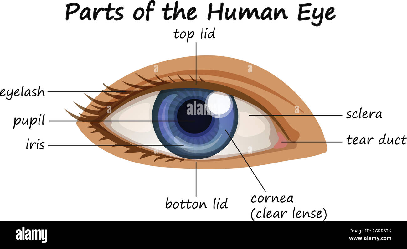
Eye Anatomy: 16 Parts of the Eye & Their Functions - Vision Center Eye Anatomy (16 Parts of the Eye & What They Do) The following are parts of the human eyes and their functions: 1. Conjunctiva The conjunctiva is the membrane covering the sclera (white portion of your eye). The conjunctiva also covers the interior of your eyelids.
How Does the Eye Work?
Eye Anatomy: Parts of the Eye and How We See - American ... Apr 29, 2023 · Eye Anatomy: Parts of the Eye Outside the Eyeball The eye sits in a protective bony socket called the orbit. Six extraocular muscles in the orbit are attached to the eye. These muscles move the eye up and down, side to side, and rotate the eye. The extraocular muscles are attached to the white part of the eye called the sclera.

Eye Anatomy | glaucoma.org
Eye Anatomy - Exeter Eye Vision occurs when light enters the eye through the pupil. With help from other important ... Exeter Eye Anatomy of the human eye diagram. Did You Know?
How the Eyes Work | National Eye Institute
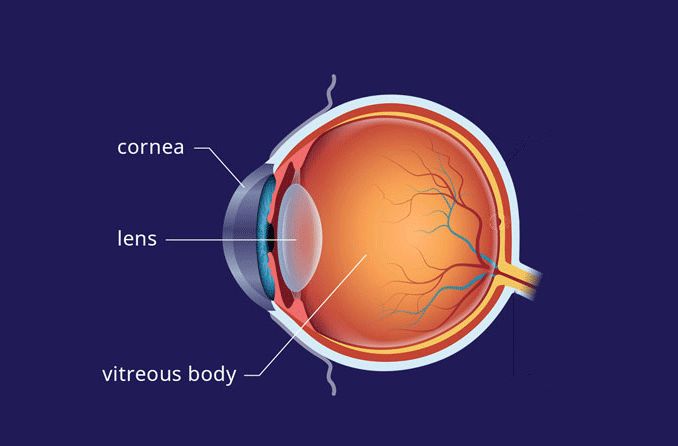
Anatomy of the Human Eye - News-Medical.net Its function is of a primary filter, before passing on the light to the lens and retina. The central portion of the front of the eyeball is termed as iris. Iris is a pigmented structure. Eye color ...

Anatomy of the Human Eye
Label Parts of the Human Eye - University of Dayton Label Parts of the Human Eye. Parts of the Eye. Select the correct label for each part of the eye. The image is taken from above the left eye. Click on the Score button to see how you did. Incorrect answers will be marked in red.
Vision and Eye Diagram: How We See
Eye Diagram With Labels and detailed description - BYJU'S Diagram Of Eye The human eye is responsible for the most important function of the human body, the sense of sight. It consists of several distinct parts that work in coordination with each other. The most common eye diseases include myopia, hypermetropia, glaucoma and cataract.

Eye Anatomy Parts & Functions | What are the Parts of the Eye ...
Eye Anatomy: A Closer Look at the Parts of the Eye When surveyed about the five senses — sight, hearing, taste, smell and touch — people consistently report that their eyesight is the mode of perception they value (and fear losing) most. Despite this, many people don’t have a good understanding of the anatomy of the eye, how visionworks or health problems that can affect the eye. Read on for a basi...

Anatomy of the eye - Moorfields Eye Hospital
The Eye - Science Quiz - Seterra - GeoGuessr The anatomy of the eye is fascinating, and this quiz game will help you ... Light enters our eyes through the pupil, then passes through a lens and the ...
Eye diagram parts hi-res stock photography and images - Alamy
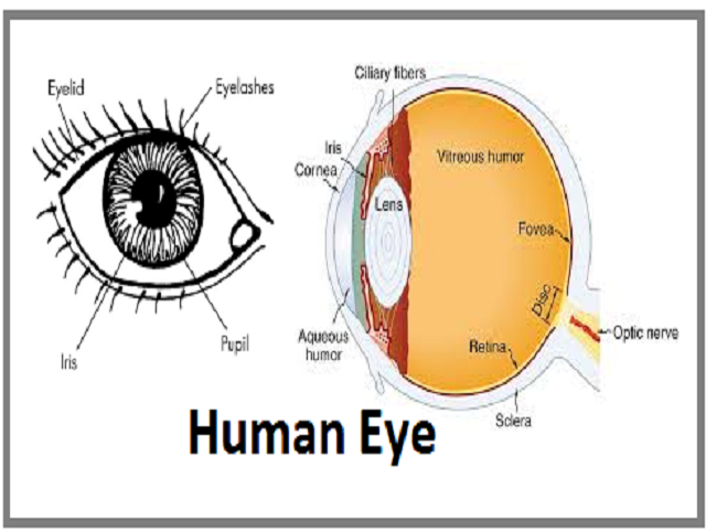
Human Eye Diagram, How The Eye Work -15 Amazing Facts of Eye The tear gland located above each eyeball and inside your upper eyelid. This gland is responsible for making a fluid that is mostly salt and water (tears) to keep the surface of your eyeball clean and moist. It also protects your eye from damage. Also Read: Brain Facts | Blood Fact s | Respiratory System | Digestive System

Human Eye Diagram, How The Eye Work -15 Amazing Facts of Eye
Eye Anatomy: The 9 Main Parts of the Eye | Specialty Eye Institute Optic Nerve. The second cranial nerve. The next component of the eye to be familiar with when you are analyzing eye anatomy is the optic nerve. The largest sensory nerve of the eye; carries impulses for sight from the retina to the brain. Composed of retinal nerve fibers that exit the eyeball through the optic disc, traverse the orbit, and pass ...

Human eye | Definition, Anatomy, Diagram, Function, & Facts ...
Structure and Function of the Eyes - Eye Disorders - MSD ...

Labelled Diagram Of Human Eye , Png Download - Label A Human ...
Problem 9, Chapter 38: Biology

4,500+ Eye Diagram Stock Photos, Pictures & Royalty-Free ...
6.831 L19: Color Design

Interactive Human Eye Anatomy

Diagram Mata Manusia Ilustrasi Stok - Unduh Gambar Sekarang ...
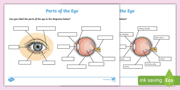
FREE! - Label the Eye Worksheet – Teacher-Made Learning Resources

Eye Diagram Quiz - ProProfs Quiz
4,500+ Eye Diagram Stock Photos, Pictures & Royalty-Free ...

Human eye Diagram Eye pattern, Eye, people, color, sports ...

Good Nutrition Can Help Your Eyes | Eye anatomy, Eye anatomy ...
Lens of the Eye - All About Vision
Eye Anatomy - Exeter Eye

Eye anatomy stock image. Image of sight, optometry, focus ...

Human eye diagram, Eye anatomy diagram, Eye anatomy

Eye - Lateral View Diagram | Quizlet

How the eye works | RNIB
Eye Diagram With Labels - ClipArt Best
How Do We See Light? | Ask A Biologist

Cow Eye Dissection & Parts of the Eye Diagram | Quizlet

Human Eye Diagram Images - Free Download on Freepik
Vision and the Structure of the Eye
Post a Comment for "39 eye diagram not labeled"