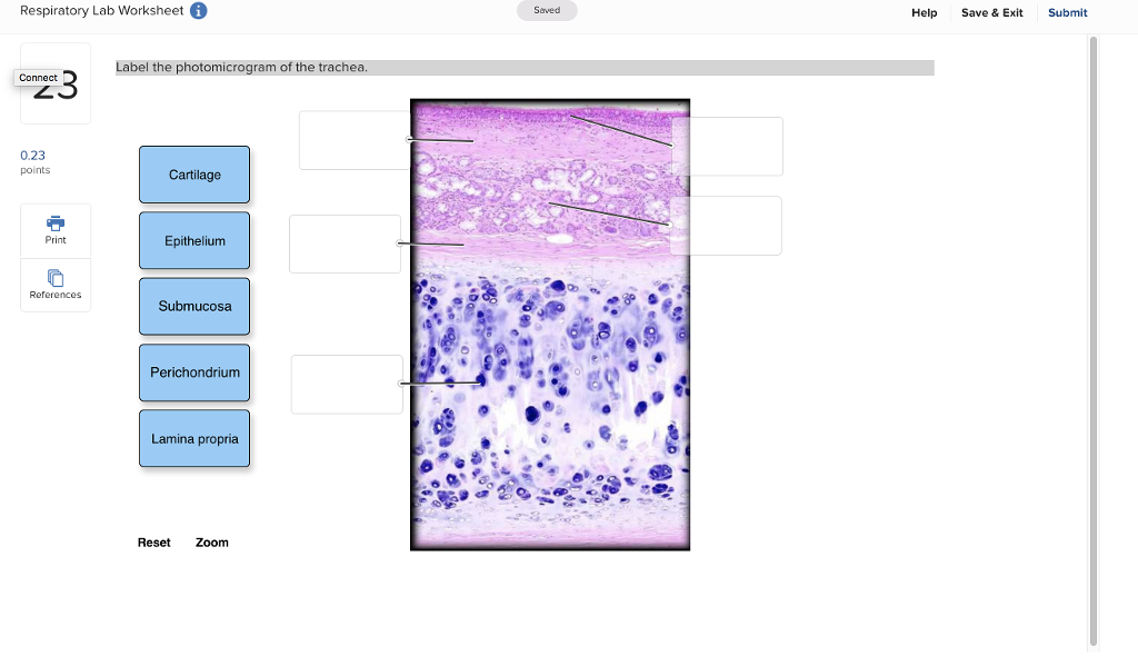
39 label the photomicrogram of the trachea
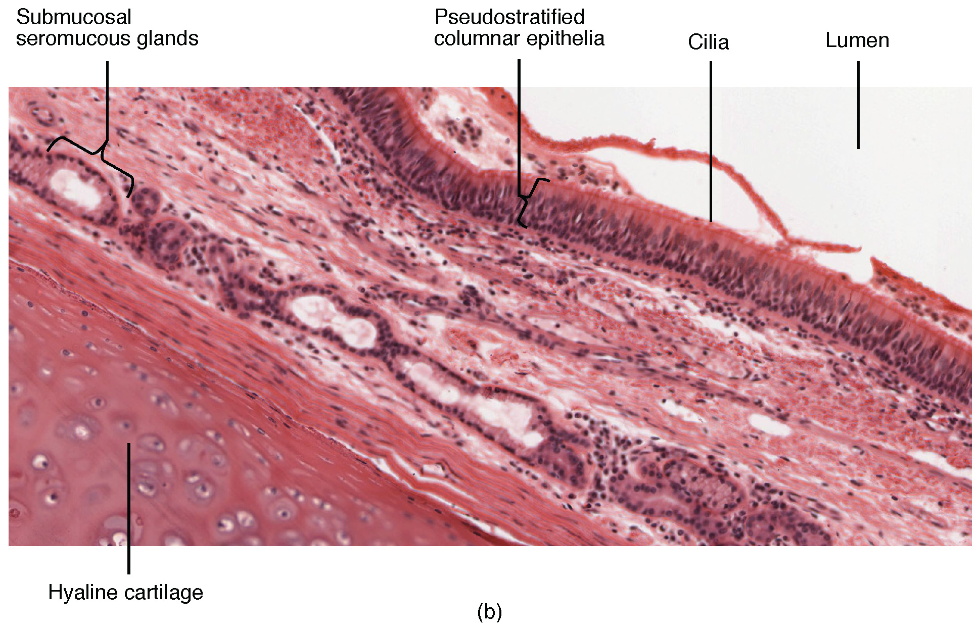
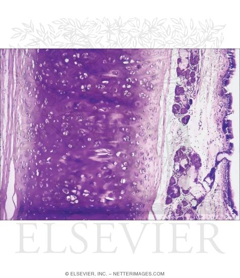
Hyaline cartilage: Histological features and cells | Kenhub Hyaline cartilage is the most common of the three types of cartilage. In its fresh state, it is homogeneous and semi-transparent. In adults, hyaline cartilage is located in the articular surfaces of movable joints, in the walls of the respiratory tracts (nose, larynx, trachea, and bronchi ), in the costal cartilages, and in the epiphyseal ... AA1 - 111.pdf - Coronary Sinus Left Diagonal Artery Right Posterior ... Label the photomicrogram of the trachea. Label the anterior view of the lower respiratory tract. Diaphragm. Put the following layers of the trachea in order ...
Bronchus and branchial wall: anatomy and diagram | GetBodySmart The bronchi are part of the airway system of the lower respiratory system. Bronchi are the branches of the trachea which provide oxygen to the lungs. In cross-section, the bronchial wall appears similar to the trachea. 1 2 Respiratory mucosa (or mucous membrane) lines the luminal surface. Mucus-secreting goblet cells are present in the epithelium.

Label the photomicrogram of the trachea
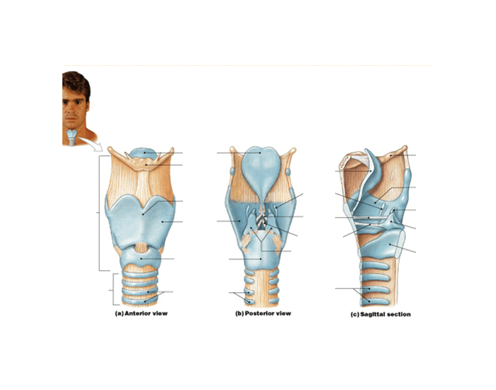
Trachea: anatomy, structure and function - GetBodySmart The outer layer of the trachea, the adventitia, is a band of loose connective tissue that loosely binds the trachea to the esophagus and other nearby organs. 1 2 A photo micrograph shows a more realistic depiction of the layers and structures that make up the tracheal wall. 1 2 An Overview of the Trachea Location, Anatomy, and Physiology: A&P 2 Lab Unit 2 Flashcards | Quizlet Label the lateral view of the larynx based on the hints if provided. Image: Label the lateral view of ... Image: Label the photomicrogram of the trachea. Photomicrograph of the tracheal wall. Panel A: showing heavy ... Photomicrograph of the tracheal wall. Panel A: showing heavy infiltration by lymphoma cells, consisting of small to mediumsized lymphocytes with plasmacytic differentiation and lymphoepithelial...
Label the photomicrogram of the trachea. Label the photomicrogram of the trachea. - Brainly.com What is the trachea? The trachea is known to be a kind of long tube that links the human larynx (voice box) to that of their bronchi. Note that the bronchi is one that send air to a person's lungs and the trachea is known to be an essential part of man's respiratory system. Trachea: Anatomy, Function, and Treatment - Verywell Health The Anatomy of the Trachea. The trachea, commonly known as the windpipe, is the large tube that delivers air from the upper respiratory tract (the nasal passages, throat, and larynx) to the bronchi (the two large airways that branch off into each lung). In the process, it warms and moisturizes the air and catches debris and microbes before they ... Photomicrograph of the trachea Diagram | Quizlet Photomicrograph of the trachea Learn Test Match Learn Test Match Created by monsth3r Terms in this set (8) Term Cilia Location Term Pseudostratified ciliated columnar Epithelium Location Term Seromucous gland Location Term Hyaline Cartilage Location Term Collagen Fibers Location Term Adventitia Location Term Submucosa Location Term Mucosa Location photomicrograph of the tracheal wall p119.1-2 .docx - In... Photomicrograph of the tracheal wallMucosa Lamina propria In submucosa End of preview Upload your study docs or become a member. View full document Students also studied 1. Sagittal eye plaque_L & UL.pdf 4 leukocytes.png 1 Trachea_with Goblet cells (p119.1-2).docx 1 Artery and Vein_Histology_L.pdf 1 Heart_Posterior (p102.1-2).docx 1
Solved Label the photomicrogram of the trachea. Cilia Lamina - Chegg Label the photomicrogram of the trachea. Cilia Lamina propria Submucosa Cilia Basement membrane Submucosa Epithelium Basement membrane Lamina propria Epithellum This problem has been solved! You'll get a detailed solution from a subject matter expert that helps you learn core concepts. See Answer Question: Label the photomicrogram of the trachea. A&P 2 Lab Unit 2 Flashcards - Quizlet Label the lateral view of the larynx based on the hints if provided. Image: Label the lateral view of ... Image: Label the photomicrogram of the trachea. Picture of the Trachea - WebMD The trachea, commonly known as the windpipe, is a tube about 4 inches long and less than an inch in diameter in most people. The trachea begins just under the larynx (voice box) and runs down... Trachea: Anatomy, blood supply, innervation and function - Kenhub Trachea. 1/5. The trachea, or windpipe, is a 10-11 cm long fibrocartilaginous tube of the lower respiratory tract. It forms the trunk of the tracheobronchial tree, or pulmonary conducting zone. The trachea extends between the larynx and thorax, consisting of two parts; cervical and thoracic. It ends at the level of the sternal angle (T5) where ...
A&P 2 Lab Unit 2 Flashcards | Quizlet The tracheal cartilage are 13-15 C-shaped cartilage rings. Identify the conchae, meatuses, vestibule, and nasopharynx. Label the photomicrogram of the lung. Identify the cartilaginous anatomical structures shown in the posterior view of the superior portion of the lower respiratory system. Photomicrograph of the tracheal wall. Panel A: showing heavy ... Photomicrograph of the tracheal wall. Panel A: showing heavy infiltration by lymphoma cells, consisting of small to mediumsized lymphocytes with plasmacytic differentiation and lymphoepithelial... A&P 2 Lab Unit 2 Flashcards | Quizlet Label the lateral view of the larynx based on the hints if provided. Image: Label the lateral view of ... Image: Label the photomicrogram of the trachea. Trachea: anatomy, structure and function - GetBodySmart The outer layer of the trachea, the adventitia, is a band of loose connective tissue that loosely binds the trachea to the esophagus and other nearby organs. 1 2 A photo micrograph shows a more realistic depiction of the layers and structures that make up the tracheal wall. 1 2 An Overview of the Trachea Location, Anatomy, and Physiology:
Practical 2 Flashcards | Quizlet
BreadAndButter/space-dict.txt at master · DreamSea ...

Histology of Respiratory System. - ppt video online download

Photomicrograph of trachea of rabbit after 3 weeks ...

Chol Mayar Anai (cholmayar16) - Profile | Pinterest

The Respiratory System: Part A - ppt download

Photomicrograph of trachea of control rabbit shows the ...
SOLVED: why is it telling me that those are wrong? Label the ...

A&P 2 Lab Unit 2 Flashcards | Quizlet

Solved 10 General Structure of Mucosa Label the structures ...

English Words | PDF
OpenStax AnatPhys fig.22.9(b) - The Trachea - English labels ...
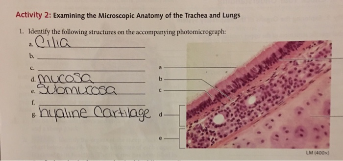
Solved 2- 1. Identify the following structures on the | Chegg.com

A&P 2 Lab Practical Final Flashcards | Quizlet

Anatomy 2 Final Lab Practical review Flashcards | Quizlet
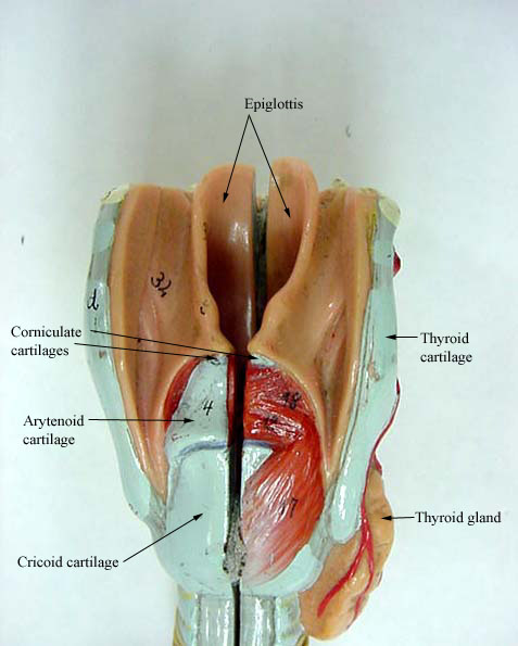
LARYNX AND TRACHEA

cartilage lab / lecture pictures Flashcards | Quizlet

A&P 2 Lab Practical Final Flashcards | Quizlet

UNIT 1 HW & Quizzes Flashcards | Quizlet
Light Micrograph of the Wall of the Trachea In Transverse Section

Respiratory system Trachea Anatomy Function Physiology, nose ...
Solved Respiratory Lab Worksheet Saved Help Save & Exit ...

Photomicrograph of trachea of rabbit after 6 weeks ...

A&P 2 Lab Unit 2 Flashcards | Quizlet

Lab Practical BIO234 -anatomy portion respratory Flashcards ...

Photomicrograph of thymic mass and trachea. The trachea ...

A&P 2 Lab Unit 2 Flashcards | Quizlet

LUNG Flashcards | Quizlet

A&P 2 Lab Unit 2 Flashcards | Quizlet

LUNG Flashcards | Quizlet

A&P 2 Lab Unit 2 Flashcards | Quizlet
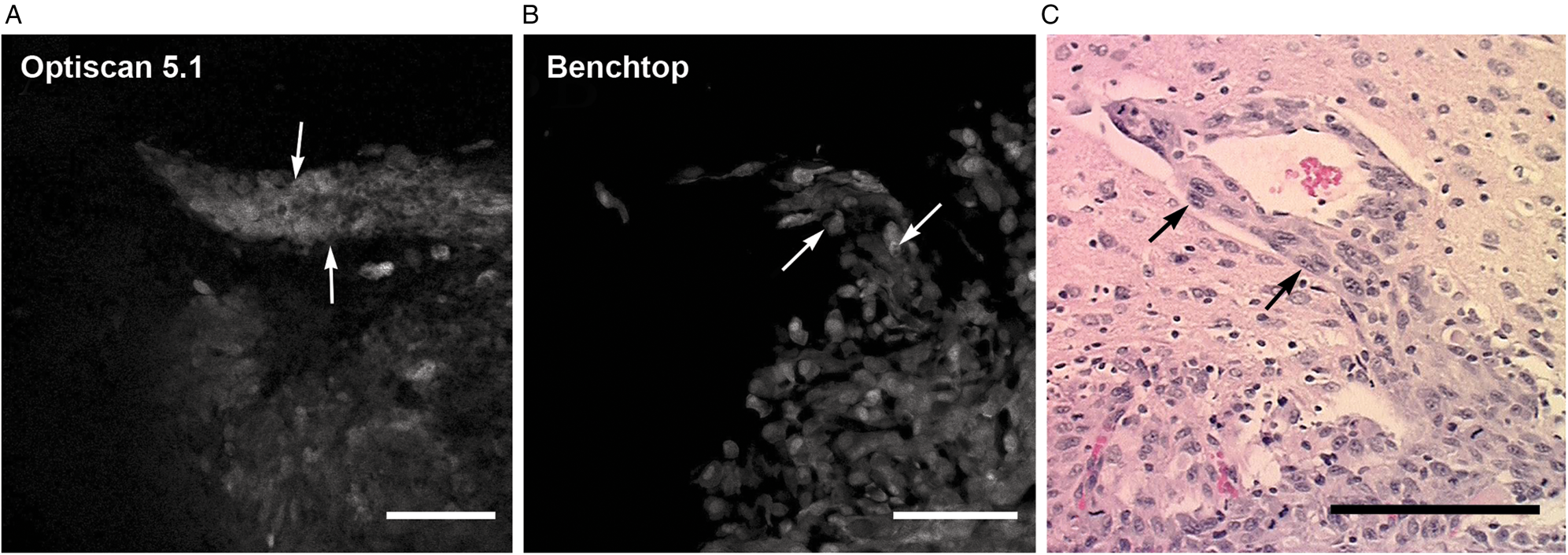
Fluorescence In Vivo Endomicroscopy Part 2: Applications of ...

Biology 234 ~ Lab MIDTERM Practical Flashcards | Quizlet
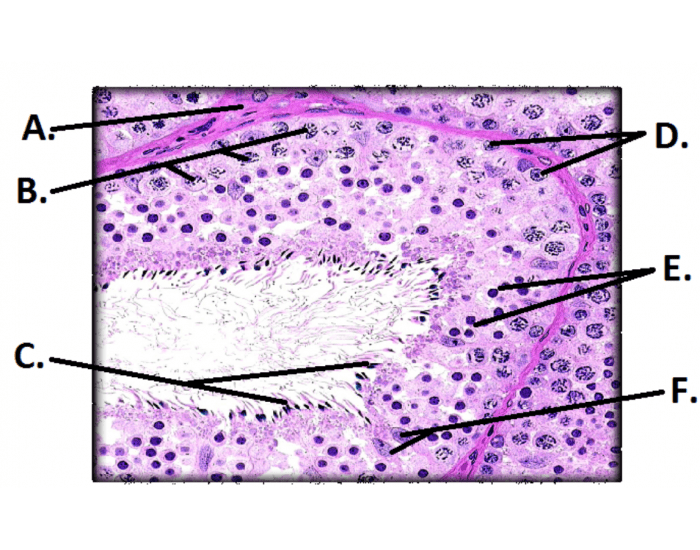
Histology of Seminiferous Tubule Quiz
Larynx & Trachea - Labeling Quiz
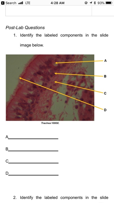
Solved Search . LTE 4:28 AM Post-Lab Questions 1. Identify ...
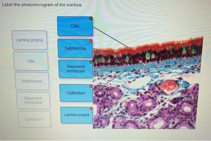
Solved Label the photomicrogram of the trachea. Cilia Lamina ...

A&P 2 Lab Practical Final Flashcards | Quizlet

A&P 2 Lab Practical Final Flashcards | Quizlet
Post a Comment for "39 label the photomicrogram of the trachea"